Understand the stages of chronic venous disease, from early venous reflux and spider veins to varicose veins with swelling, skin changes and venous ulcers.
Some people remain symptom-free for years, while others develop pain, swelling, skin changes or ulcers. Early vascular assessment can help identify the underlying venous problem.
Varicose veins are enlarged, twisted superficial veins, usually affecting the lower limbs. They develop when vein walls and valves do not function properly, allowing blood to flow backward toward the feet and increasing pressure within the veins.
The leg has superficial veins, deep veins and perforating veins that connect the superficial and deep systems. Normally, venous valves open to allow blood to move toward the heart and close to prevent backward flow. When valves leak, venous reflux can cause pooling of blood and venous hypertension.
The source page describes the clinical component of the C-E-A-P classification for chronic venous disease. C refers to clinical signs, E to etiology, A to anatomic distribution and P to pathophysiology.
Venous reflux may be present, but there are no visible clinical signs of venous disease.
Small visible superficial veins, including spider veins or telangiectasia, may be present.
Visible enlarged or tortuous varicose veins are present.
Varicose veins are associated with swelling, commonly around the ankle or lower leg.
More advanced chronic venous disease may cause skin changes such as discoloration, dermatitis or other tissue changes.
C5 represents a healed venous ulcer, while C6 represents an active venous ulcer. Advanced C4–C6 disease is commonly described as chronic venous insufficiency.
The source page notes that symptoms may include pain or aching, burning, itching, numbness, heaviness and ankle swelling. Symptoms may be worse toward the end of the day or in warm weather.
Aching or discomfort around visible veins may occur, particularly after prolonged standing.
A heavy or tired feeling in the calf can accompany chronic venous reflux.
Oedema around the ankles or lower legs can occur as venous disease progresses.
Burning, itching or skin irritation may accompany symptomatic varicose veins.
More advanced venous disease can cause reddish, brown or dark skin changes around the lower leg.
Advanced disease may be associated with non-healing venous ulcers or bleeding from varicose veins.
Varicose veins may be primary, secondary or associated with congenital venous abnormalities. Several factors can increase symptoms or risk.
Weak or damaged valves can allow blood to reflux toward the feet, increasing pressure in the superficial veins.
Genetic predisposition can contribute to the development of varicose veins.
Pregnancy can aggravate varicose veins in people with a predisposition. The source page notes that veins often improve after delivery, although they may persist after repeated pregnancies.
Occupations requiring long periods of standing can aggravate symptoms and venous pressure.
The source page identifies obesity as an aggravating factor for varicose vein symptoms.
Secondary varicose veins can occur when superficial venous reflux is associated with obstruction of the deep veins, including after previous deep vein thrombosis.
The source page states that diagnosis can often be made by a vascular surgeon through physical examination and Doppler assessment, with duplex ultrasonography used when additional information is needed.
The vascular specialist examines the visible veins, swelling, skin changes and other clinical findings.
Continuous-wave Doppler can help assess venous blood flow during clinical evaluation.
Duplex ultrasound can assess venous reflux and help determine the anatomy and treatment approach when indicated.
Treatment depends on symptoms, venous anatomy, the stage of chronic venous disease and the findings on vascular assessment.
Graduated compression stockings may be recommended for symptom relief or as part of management. The appropriate compression level should be selected with medical advice.
Endovenous laser therapy uses thermal energy to ablate an affected saphenous vein while leaving it in place.
Radiofrequency treatment uses controlled thermal energy to close an affected vein and redirect blood through healthier venous pathways.
Ultrasound-guided foam sclerotherapy can be used for selected veins. The source page notes that indications and results can vary.
Selected patients may be treated using a non-thermal adhesive technique to close the diseased vein.
In selected cases, damaged veins or their connections may be surgically removed or treated. The specific procedure depends on the venous anatomy and clinical findings.
The source page also discusses pelvic varices associated with reflux in ovarian or internal iliac veins. These can produce varicose veins around the vagina, vulva or upper medial thigh.
When pelvic varices persist and are clinically appropriate for treatment, the source page describes venography and embolization of responsible or feeding veins, sometimes combined with local avulsions or sclerotherapy.
Reduce long periods of standing where possible and take short walking breaks.
If sitting for long periods, walk for a few minutes or regularly flex the legs.
When practical, elevate your legs above heart level for periods of rest.
Use compression stockings only as advised by your doctor and follow the recommended fit and compression level.
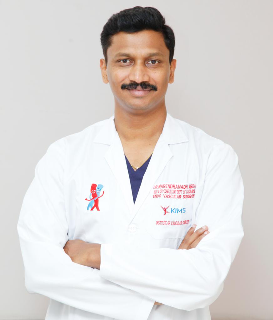
Consultant Vascular & Endovascular Surgeon
Dr. Narendranadh Meda is a vascular and endovascular surgeon associated with Hyderabad Vascular Center, providing evaluation and treatment for arterial and venous vascular conditions.
Varicose vein treatment starts with understanding the venous anatomy and identifying reflux or obstruction. Depending on the findings and stage of disease, management may include compression, endovenous treatment, sclerotherapy or surgery.
Book a ConsultationThe clinical C-E-A-P stages range from C0 with no visible signs, C1 spider veins, C2 varicose veins, C3 varicose veins with oedema, C4 skin changes, C5 healed ulcer and C6 active venous ulcer.
Yes. The source page states that leg varicose veins tend to get worse with time, although the rate of progression varies between people.
No. Some people remain symptom-free for a long period. Treatment depends on symptoms, complications, venous anatomy and the findings of vascular assessment.
Physical examination and Doppler assessment may be sufficient in some cases. Duplex ultrasound can provide additional information when required.
Depending on the patient, options include compression therapy, EVLT, radiofrequency ablation, selected foam sclerotherapy, medical adhesive treatment and surgical treatment.
Yes. The source page notes that new varicose veins may develop after a successful procedure over time, even though the veins already treated may remain closed or removed.
Get a vascular assessment to understand the stage of venous disease and whether observation, compression or a definitive treatment may be appropriate.
